

Crystal characteristics of fibrous calcite veins based on Electron Back Scattered Diffraction (EBSD)
-
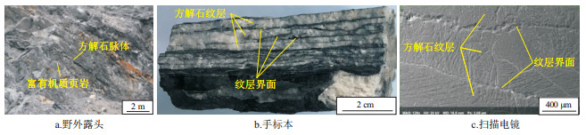
摘要: 方解石脉体广泛发育在富有机质页岩中,其岩石学特征和成因机制是研究的热点。电子背散射衍射技术能够原位表征矿物晶体的微观结构和晶体取向信息,而被广泛应用于材料科学领域,并在地质学领域得到了迅速发展。为了明确四川盆地龙马溪组富有机质页岩中纤维状方解石脉体的晶体特征,电子背散射衍射技术被用于表征方解石脉体的矿物学特征和结晶学特征。研究结果表明,方解石脉体的矿物组成为方解石和石英,其中方解石是主体,其晶粒的平均大小为372 μm;而石英主要分布在方解石纹层界面处。方解石脉体中的方解石晶体属于三方晶系或菱方晶系,相应的晶胞类型为三方晶胞或菱方晶胞,其晶格常数为a0=b0=4.99 Å,c0=17.061 Å,α=β=90°,γ=120°。方解石脉体在纵剖面上具有一定的择优取向,原因是方解石晶粒内部发育聚片双晶,其中相邻的双晶条纹具有不同的晶体取向,晶体取向差为75°;而相间的双晶条纹具有相同的晶体取向,且同一双晶条纹的晶体取向相同。方解石晶粒内部发育完全解理,常成组出现且与双晶条纹呈锐夹角斜交,两者均是在方解石结晶过程中受到构造压扭作用产生的,其中最大主应力方向与双晶条纹平行。Abstract: Calcite veins are widely developed in organic-rich shale, and their petrological characteristics and genetic mechanism are the focus of research. The Electron Back Scatter Diffraction (EBSD) can characterize the micro-structure and orientation of mineral crystals in situ, which has been widely used in the field of material science and has been rapidly developed in the field of geology. In order to clarify the crystal characte-ristics of fibrous calcite veins in organic-rich shale of the Longmaxi Formation in the Sichuan Basin, EBSD was used to characterize the mineralogical and crystallographic characteristics of calcite veins. The calcite veins are mainly composed of calcite and quartz. Calcite is the main body, with an average grain size of 372 μm, while quartz is mainly distributed at the interface of calcite lamina. The calcite crystals in calcite veins belong to trigonal[JP] or rhombohedral system, and the corresponding unit cell is trigonal or rhombohedral. The lattice parameters are a0=b0=4.99 Å, c0=17.061 Å, α=β=90°, γ=120°, respectively. Calcite veins have a certain preferred orientation on longitudinal section, which is due to the development of polysynthetic twin crystals in calcite grains. The adjacent twin crystal stripes have different crystal orientations, and the crystal misorientation is 75° while the alternate twin stripes have the same crystal orientation, and the crystal orientation of the same twin stripe is the same. In calcite grains, perfect cleavage occurs in groups with sharp angle with twin crystal stripes. Both cleavage and twin crystal stripes are formed by tectonic compression and shearing during the crystallization of calcite, and the maximum principal stress direction is parallel to the twin crystal stripes.
-

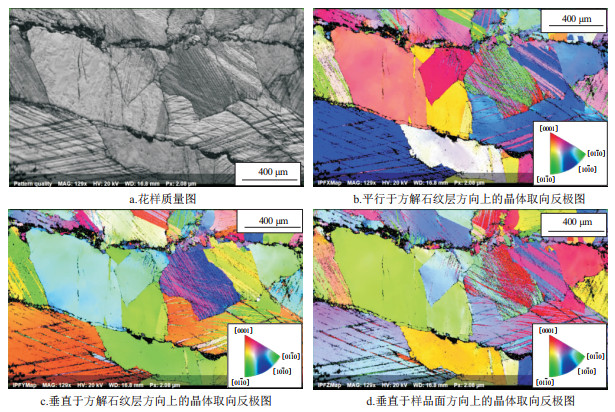
图 4 方解石脉体花样质量图及反极图
Figure 4. Pattern quality figure and inverse pole figure of calcite veins

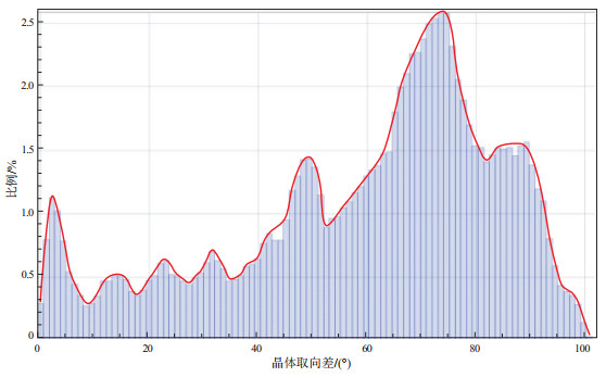
图 7 方解石脉体纵剖面上的晶体取向差分布
Figure 7. Distribution of crystal misorientation on longitudinal section of calcite veins

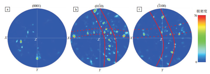
图 8 方解石脉体纵剖面上的晶体极图
Figure 8. Crystal pole figure on longitudinal section of calcite veins

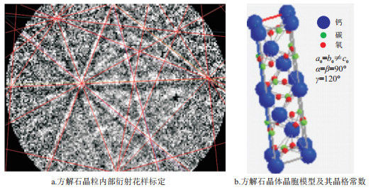
图 9 方解石晶粒内部电子背散射衍射花样标定及晶胞提取
Figure 9. Calibration of EBSD pattern and extraction of crystal cell within calcite grains

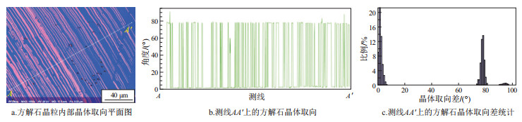
图 10 方解石晶粒内部双晶及其晶体取向
Figure 10. Twin crystals and crystal orientation within calcite grains
-
[1] 王冠民, 任拥军, 钟建华, 等. 济阳坳陷古近系黑色页岩中纹层状方解石脉的成因探讨[J]. 地质学报, 2005, 79(6): 834-838. doi: 10.3321/j.issn:0001-5717.2005.06.012WANG Guanmin, REN Yongjun, ZHONG Jianhua, et al. Genetic analysis on lamellar calcite veins in Paleogene black shale of the Jiyang Depression[J]. Acta Geologica Sinica, 2005, 79(6): 834-838. doi: 10.3321/j.issn:0001-5717.2005.06.012 [2] ZHANG Jianguo, JIANG Zaixing, JIANG Xiaolong, et al. Oil generation induces sparry calcite formation in lacustrine mudrock, Eocene of East China[J]. Marine and Petroleum Geo-logy, 2016, 71: 344-359. doi: 10.1016/j.marpetgeo.2016.01.007 [3] TABER S. The origin of veinlets in the Silurian and Devonian strata of central New York[J]. The Journal of Geology, 1918, 26(1): 56-73. doi: 10.1086/622561 [4] BONS P D, MONTENARI M. The formation of antitaxial calcite veins with well-developed fibres, Oppaminda Creek, South Australia[J]. Journal of Structural Geology, 2005, 27(2): 231-248. doi: 10.1016/j.jsg.2004.08.009 [5] PUTNIS A, PRIETO M, FERNANDEZ-DIAZ L. Fluid supersaturation and crystallization in porous media[J]. Geological Magazine, 1995, 132(1): 1-13. doi: 10.1017/S0016756800011389 [6] COBBOLD P R, ZANELLA A, RODRIGUES N, et al. Bedding-parallel fibrous veins (beef and cone-in-cone): worldwide occurrence and possible significance in terms of fluid overpressure, hydrocarbon generation and mineralization[J]. Marine and Petroleum Geology, 2013, 43: 1-20. doi: 10.1016/j.marpetgeo.2013.01.010 [7] BONS P D, JESSELL M W. Experimental simulation of the formation of fibrous veins by localised dissolution-precipitation creep[J]. Mineralogical Magazine, 1997, 61(404): 53-63. doi: 10.1180/minmag.1997.061.404.06 [8] MAHER H D, OGATA K, BRAATHEN A. Cone-in-cone and beef mineralization associated with Triassic growth basin faulting and shallow shale diagenesis, EdgeØya, Svalbard[J]. Geological Magazine, 2017, 154(2): 201-216. doi: 10.1017/S0016756815000886 [9] MENG Qingfeng, HOOKER J, CARTWRIGHT J. Early overpressuring in organic-rich shales during burial: evidence from fibrous calcite veins in the Lower Jurassic Shales-with-Beef Member in the Wessex Basin, UK[J]. Journal of the Geological Society, 2017, 174(5): 869-882. doi: 10.1144/jgs2016-146 [10] UKAR E, LOPEZ R G, GALE J F W, et al. New type of kinematic indicator in bed-parallel veins, Late Jurassic-Early Cretaceous Vaca Muerta Formation, Argentina: E-W shortening during Late Cretaceous vein opening[J]. Journal of Structural Geology, 2017, 104: 31-47. doi: 10.1016/j.jsg.2017.09.014 [11] WANG Miao, CHEN Yong, BAIN W M, et al. Direct evidence for fluid overpressure during hydrocarbon generation and expulsion from organic-rich shales[J]. Geology, 2020, 48(4): 374-378. doi: 10.1130/G46650.1 [12] MA Cunfei, DONG Chunmei, LUAN Guoqiang, et al. Types, characteristics and effects of natural fluid pressure fractures in shale: a case study of the Paleogene strata in Eastern China[J]. Petroleum Exploration and Development, 2016, 43(4): 634-643. doi: 10.1016/S1876-3804(16)30074-X [13] LUAN Guoqiang, DONG Chunmei, AZMY K, et al. Origin of bedding-parallel fibrous calcite veins in lacustrine black shale: a case study from Dongying Depression, Bohai Bay Basin[J]. Marine and Petroleum Geology, 2019, 102: 873-885. doi: 10.1016/j.marpetgeo.2019.01.010 [14] SHOVKUN I, ESPINOZA D N. Geomechanical implications of dissolution of mineralized natural fractures in shale formations[J]. Journal of Petroleum Science and Engineering, 2018, 160: 555-564. doi: 10.1016/j.petrol.2017.10.043 [15] MILLIKEN K L, DAY-STIRRAT R J. Cementation in mudrocks: brief review with examples from cratonic basin mudrocks[M]//CHATELLIER J Y, JARVIE D M. Critical assessment of shale resource plays. Tulsa: American Association of Petroleum Geo-logists, 2013: 133-160. [16] LOUCKS R G, REED R M, RUPPEL S C, et al. Spectrum of pore types and networks in mudrocks and a descriptive classification for matrix-related mudrock pores[J]. AAPG Bulletin, 2012, 96(6): 1071-1098. doi: 10.1306/08171111061 [17] GALE J F W, REED R M, HOLDER J. Natural fractures in the Barnett shale and their importance for hydraulic fracture treatments[J]. AAPG Bulletin, 2007, 91(4): 603-622. doi: 10.1306/11010606061 [18] ZHANG Bo, YIN Congyuan, GU Zhidong, et al. New indicators from bedding-parallel beef veins for the fault valve mechanism[J]. Science China Earth Sciences, 2015, 58(8): 1320-1336. doi: 10.1007/s11430-015-5086-6 [19] MING Xiaoran, LIU Li, YU Miao, et al. Bleached mudstone, iron concretions, and calcite veins: a natural analogue for the effects of reducing CO2-bearing fluids on migration and mineralization of iron, sealing properties, and composition of mudstone cap rocks[J]. Geofluids, 2016, 16(5): 1017-1042. doi: 10.1111/gfl.12203 [20] VILLERT S, MAURICE C, WYON C, et al. Accuracy assessment of elastic strain measurement by EBSD[J]. Journal of microscopy, 2009, 233(2): 290-301. doi: 10.1111/j.1365-2818.2009.03120.x [21] WILKINSON A J, MEADEN G, DINGLEY D J, et al. Mapping strains at the nanoscale using electron back scatter diffraction[J]. Superlattices and Microstructures, 2009, 45(4/5): 285-294. [22] MAURICE C, QUEY R, FORTUNIER R, et al. High angular resolution EBSD and its materials applications[M]//MOLODOV D A. Microstructural design of advanced engineering materials. Weinheim: Wiley-VCH Verlag GmbH & Co. KGaA, 2013: 339-365. [23] 黄亚敏, 潘春旭. 基于电子背散射衍射(EBSD)技术的材料微区应力应变状态研究综述[J]. 电子显微学报, 2010, 29(1): 1-11. https://www.cnki.com.cn/Article/CJFDTOTAL-DZXV201001002.htmHUANG Yamin, PAN Chunxu. Micro-stress-strain analysis in materials based upon EBSD technique: a review[J]. Journal of Chinese Electron Microscopy Society, 2010, 29(1): 1-11. https://www.cnki.com.cn/Article/CJFDTOTAL-DZXV201001002.htm [24] 杨平. 电子背散射衍射技术、几何晶体学与材料科学[J]. 电子显微学报, 2008, 27(6): 425-431. doi: 10.3969/j.issn.1000-6281.2008.06.001YANG Ping. EBSD technique, geometric crystallography and materials science[J]. Journal of Chinese Electron Microscopy Society, 2008, 27(6): 425-431. doi: 10.3969/j.issn.1000-6281.2008.06.001 [25] 靳丽, MISHARA R K, KUBIC R. 材料变形过程中的原位电子背散射衍射(in-situ EBSD)分析[J]. 电子显微学报, 2008, 27(6): 439-442. https://www.cnki.com.cn/Article/CJFDTOTAL-QHJJ202202007.htmJIN Li, MISHARA R K, KUBIC R. In-situ EBSD analysis on the microstructures during deformation[J]. Journal of Chinese Electron Microscopy Society, 2008, 27(6): 439-442. https://www.cnki.com.cn/Article/CJFDTOTAL-QHJJ202202007.htm [26] NOWELL M M, WRIGHT S L. Phase differentiation via combined EBSD and XEDS[J]. Journal of Microscopy, 2005, 213(3): 296-305. [27] ASPIROZ M D, LLOYD G E, FERNÁNDEZ C. Development of lattice preferred orientation in clinoamphiboles deformed under low-pressure metamorphic conditions. A SEM/EBSD study of metabasites from the Aracena metamorphic belt (SW Spain)[J]. Journal of Structural Geology, 2007, 29(4): 629-645. doi: 10.1016/j.jsg.2006.10.010 [28] ZAEFFERER S, WRIGHT S L. 3D characterization of crystallographic orientation in polycrystals via EBSD[J]. Chinese Journal of Stereology and Image Analysis, 2007, 12(4): 233-238. [29] 刘俊来, 曹淑云, 邹运鑫, 等. 岩石电子背散射衍射(EBSD)组构分析及应用[J]. 地质通报, 2008, 27(10): 1638-1645. https://www.cnki.com.cn/Article/CJFDTOTAL-ZQYD200810006.htmLIU Junlai, CAO Shuyun, ZOU Yunxin, et al. EBSD analysis of rock fabrics and its application[J]. Geological Bulletin of China, 2008, 27(10): 1638-1645. https://www.cnki.com.cn/Article/CJFDTOTAL-ZQYD200810006.htm [30] HEIDELBACH F, KUNZE K, WENK H R. Texture analysis of a recrystallized quartzite using electron diffraction in the scanning electron microscope[J]. Journal of Structural Geology, 2000, 22(1): 91-104. doi: 10.1016/S0191-8141(99)00125-X [31] LLOYD G E. Grain boundary contact effects during faulting of quartzite: an SEM/EBSD analysis[J]. Journal of Structural Geology, 2000, 22(11/12): 1675-1693. [32] MAINPRICE D, BASCOU J, CORDIER P, et al. Crystal preferred orientations of garnet: comparison between numerical simulations and electron back-scattered diffraction (EBSD) measurements in naturally deformed eclogites[J]. Journal of Structural Geology, 2004, 26(11): 2089-2102. doi: 10.1016/j.jsg.2004.04.008 [33] TOY V G, PRIOR D J, NORRIS R J. Quartz fabrics in the Alpine Fault mylonites: influence of pre-existing preferred orientations on fabric development during progressive uplift[J]. Journal of Structural Geology, 2008, 30(5): 602-621. doi: 10.1016/j.jsg.2008.01.001 [34] 许志琴, 王勤, 梁凤华, 等. 电子背散射衍射(EBSD)技术在大陆动力学研究中的应用[J]. 岩石学报, 2009, 25(7): 1721-1736. https://www.cnki.com.cn/Article/CJFDTOTAL-YSXB200907016.htmXU Zhiqin, WANG Qin, LIANG Fenghua, et al. Electron backscatter diffraction (EBSD) technique and its application to study of continental dynamics[J]. Acta Petrologica Sinica, 2009, 25(7): 1721-1736. https://www.cnki.com.cn/Article/CJFDTOTAL-YSXB200907016.htm [35] MICHAEL J, SCHISCHKA J, ALTMANN F. HKL technology EBSD application catalogue[M]. Hobro, Denmark: HKL Technology. 2003. [36] BURKHARD M. Calcite twins, their geometry, appearance and significance as stress-strain markers and indicators of tectonic regime: a review[J]. Journal of Structural Geology, 1993, 15(3/5): 351-368. [37] 陈世悦. 矿物岩石学[M]. 东营: 中国石油大学出版社, 2002.CHEN Shiyue. Mineralogy petrology[M]. Dongying: China University of Petroleum Press, 2002. [38] 戴俊生. 构造地质学及大地构造[M]. 北京: 石油工业出版社, 2006.DAI Junsheng. Structural geology and tectonics[M]. Beijing: Petroleum Industry Press, 2006. [39] 董大忠, 施振生, 孙莎莎, 等. 黑色页岩微裂缝发育控制因素: 以长宁双河剖面五峰组—龙马溪组为例[J]. 石油勘探与开发, 2018, 45(5): 763-774. https://www.cnki.com.cn/Article/CJFDTOTAL-SKYK201805003.htmDONG Dazhong, SHI Zhensheng, SUN Shasha, et al. Factors contro-lling microfractures in black shale: a case study of Ordovician Wufeng Formation Silurian Longmaxi Formation in Shuanghe profile, Changning area, Sichuan Basin, SW China[J]. PetroleumExploration and Development, 2018, 45(5): 763-774. https://www.cnki.com.cn/Article/CJFDTOTAL-SKYK201805003.htm [40] 梁峰, 邱峋晰, 戴赟, 等. 四川盆地下志留统龙马溪组页岩纳米孔隙发育特征及主控因素[J]. 石油实验地质, 2020, 42(3): 451-458. doi: 10.11781/sysydz202003451LIANG Feng, QIU Xunxi, DAI Yun, et al. Characteristics and main controls of nano-pores in the Lower Silurian Longmaxi shale, Sichuan Basin[J]. Petroleum Geology & Experiment, 2020, 42(3): 451-458. doi: 10.11781/sysydz202003451 -

 下载:
下载:


 点击查看大图
计量
- 文章访问数: 403
- HTML全文浏览量: 122
- PDF下载量: 43
- 被引次数: 0
 苏公网安备32021102000780号