

Discussion on key technologies in micro-CT experiments and their applications in oil and gas exploration
-
摘要: 微米CT技术在油气田勘探与开发过程中已得到广泛应用,但关键控制参数、数据处理方法等环节尚未形成统一的规范,其数据的准确性和可比性仍受到较大影响。为系统研究微米CT实验参数对测试结果的影响,选取南海北部湾盆地和莺歌海盆地的7块典型样品,重点探讨检测分辨率、表征性体积单元大小及数据处理方式对实验结果的影响。研究结果表明:(1)固定的检测分辨率是确保数据可靠性的关键因素,对孔隙结构参数的提取及后续分析影响最为显著,应综合考虑样品尺寸和岩性特征等因素进行优化设置;(2)在表征砂岩样品孔隙度时,较大的体积单元能提高结果的准确性,而在构建孔隙网络模型时,表征性体积单元应不小于600体素×600体素×600体素,以确保网络模型的代表性;(3)采用分区间统计法计算孔隙直径(Φ)的累积体积频率,并绘制孔径分布曲线,同时使用体积法计算平均孔径,可更精确地表征岩石的孔隙结构特征。研究结果不仅为微米CT技术在油气勘探中的测试流程和应用提供了理论支撑,同时也为实验规范的制定提供了参考。Abstract: Micro-CT technology has been widely applied in oil and gas exploration and development. However, unified standards for key control parameters and data processing methods have yet to be established, significantly affecting data accuracy and comparability. To systematically study the impact of micro-CT experimental parameters on test results, seven representative samples from the Beibuwan Basin and the Yinggehai Basin in the South China Sea were used as the research subjects. The research focused on the impact of scanning resolution, representative volume element (RVE) size, and data processing methods on experimental outcomes. The findings indicated that: (1) A fixed scanning resolution is a key factor in ensuring data reliability, as it significantly affects the extraction of pore structure parameters and subsequent analysis. Resolution should be optimized considering sample size and lithological characteristics. (2) For characterizing the porosity of sandstone samples, better result accuracy can be achieved by increasing the RVE size. While constructing pore network models, the RVE size should be no smaller than 600×600×600 voxels to ensure model representativeness. (3) Use interval-based statistics to calculate the cumulative volume frequency of pore diameters (Φ), and plot the pore size distribution curves. Use the volumetric method to calculate the average pore diameter. These methods can provide a more accurate characterization of rock pore structure. The study offers theoretical support for the application and workflow optimization of micro-CT technology in oil and gas exploration, providing a reference for establishing experimental standards.
-

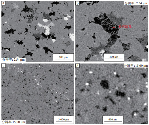
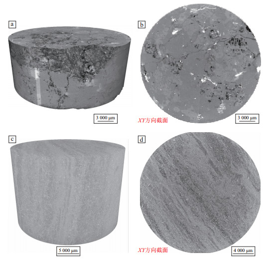
图 1 北部湾盆地1号砂岩样品不同分辨率下CT扫描XY截面灰度
Figure 1. Grayscale CT scans of XY cross-section at different resolutions for sandstone sample No. 1 from Beibuwan Basin

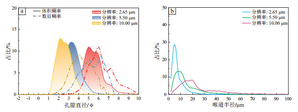
图 2 北部湾盆地1号砂岩样品不同分辨率下孔隙直径和喉道半径分布频率
Figure 2. Pore diameter and throat radius frequency distributions at different resolutions for sandstone sample No. 1 from Beibuwan Basin

图 3 北部湾盆地2号砂岩样品不同分辨率下CT扫描XY截面灰度
Figure 3. Grayscale CT scans of XY cross-section at different resolutions for sandstone sample No. 2 from Beibuwan Basin

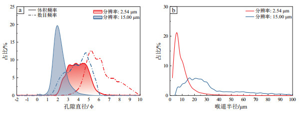
图 4 北部湾盆地2号砂岩样品不同分辨率下孔隙直径和喉道半径分布频率
Figure 4. Pore diameter and throat radius distribution frequency at different resolutions for sandstone sample No. 2 from Beibuwan Basin

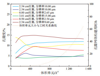
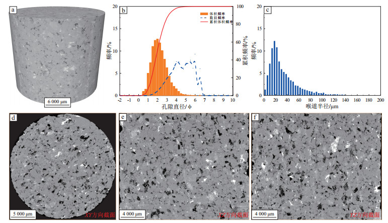
图 5 北部湾盆地和莺歌海盆地砂岩CT扫描孔隙度与孔隙网络模型构建用时随体积单元变化
Figure 5. Variation of porosity in CT scans and time for constructing pore network models with volume element in sandstones from Beibuwan and Yinggehai basins

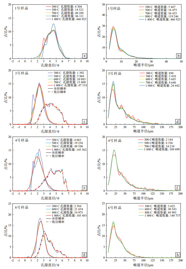
图 6 北部湾盆地1号样、3号样、4号样和莺歌海盆地6号样CT扫描孔隙及喉道分布频率
Figure 6. Pore and throat distribution frequency in CT scans of samples No. 1, 3, and 4 from Beibuwan Basin and sample No. 6 from Yinggehai Basin

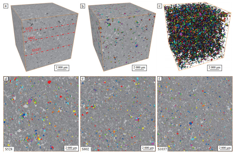
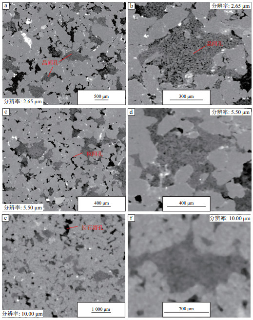
图 7 北部湾盆地3号样品CT扫描(a-c)与模拟铸体薄片(d-f)孔隙图像对比
Figure 7. Comparison of CT scans (a-c) and simulated cast thin-section (d-f) pore images of sample No. 3 from Beibuwan Basin

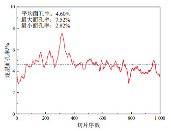
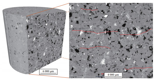
图 8 北部湾盆地3号样品CT扫描岩石逐层面孔率分布
Figure 8. Layer-by-layer pore fraction distribution of the rock in CT scans of sample No. 3 from Beibuwan Basin

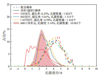
图 9 北部湾盆地3号样品CT扫描法与薄片图像法的孔隙分布频率对比
Figure 9. Comparison of pore distribution frequency between CT scanning and thin-section imaging for sample No. 3 from Beibuwan Basin

图 10 北部湾盆地5号样品(a-b)和莺歌海盆地7号样品(c-d)CT扫描3D视图及XY方向截面灰度图
Figure 10. 3D CT scans and XY cross-section grayscale of sample No. 5 from Beibuwan Basin(a-b) and sample No. 7 from Yinggehai Basin(c-d)

图 11 莺歌海盆地6号样品CT扫描灰度图及孔隙结构参数直方图
Figure 11. Grayscale CT scans and pore structure parameter histograms of sample No. 6 from Yinggehai Basin

图 12 莺歌海盆地6号样品CT扫描YZ方向截面微裂缝特征灰度
Figure 12. Microcrack characteristics in grayscale CT scans of YZ cross-section of sample No. 6 from Yinggehai Basin
表 1 北部湾盆地和莺歌海盆地实验样品信息
Table 1. Information of experimental samples from Beibuwan and Yinggehai basins
盆地 样品编号 深度/m 岩石学特征 孔隙度/% 渗透率/10-3 μm2 北部湾盆地 1 3 026.98 长石石英细—中砂岩,压实作用强,孔隙类型以长石溶孔、高岭石晶间孔为主,少量粒间孔。 16.70 0.759 2 3 548.00 长石石英中—粗砂岩,压实作用强,孔隙类型以长石溶蚀孔、铸模孔为主。 10.60 0.157 3 3 497.50 含碳酸盐不等粒砂岩,碳酸盐不均匀胶结,孔隙类型以长石溶孔、铸模孔为主。 11.70 1.344 4 3 490.00 含碳酸盐中—粗砂岩,碳酸盐不均匀胶结,孔隙类型以粒间孔为主。 21.73 3 200.147 5 1 024.26 碎裂花岗岩,花岗结构,成分主要为钾长石、石英,储集空间主要为长石溶蚀孔隙和少量裂缝。 13.20 1.712 莺歌海盆地 6 4 215.80 长石岩屑石英粗—极粗砂岩,压实作用强,岩屑含量较丰富,长石溶孔及铸模孔发育,原生粒间孔少,整体孔隙差。 11.77 1.690 7 3 170.48 泥砂互层,砂质以极细粒为主,部分粉砂,呈条带状或透镜状分布;泥质主要呈条带状分布。 8.03 0.081  下载: 导出CSV
表 2 北部湾盆地1号样品和2号样品微米CT扫描孔隙结构参数
Table 2. Micro-CT computed pore structure parameters of samples No. 1 and No. 2 from Beibuwan Basin
样品编号 样品直径/cm 分辨率/(μm /体素) 孔隙度/% 平均孔径/μm 喉道半径/μm 喉道长度/μm 配位数/个 1 2.54 10.00 5.05 177.58 25.63 137.05 0.86 1-1 0.50 5.50 7.91 109.37 13.78 75.33 1.57 1-1 0.50 2.65 9.66 25.97 6.69 37.81 2.77 2 3.81 15.00 9.80 239.30 33.95 147.92 0.76 2-1 0.50 2.54 11.47 70.88 9.23 47.80 2.39
下载: 导出CSV
表 3 北部湾盆地1号样、3号样、4号样和莺歌海盆地6号样CT扫描不同体积单元孔隙结构参数
Table 3. Pore structure parameters of different volume elements in CT scans for samples No. 1, 3, and 4 from Beibuwan Basin and sample No. 6 from Yinggehai Basin
样品编号 样品直径/cm 分辨率/(μm/体素) 类型 体积单元大小/体素 孔隙度/% 孔隙数/个 喉道数/个 体积法平均孔径/μm 数量法平均孔径/μm 平均喉道半径/μm 平均喉道长度/μm 平均配位数/个 1 0.80 2.65 三维CT 300×300×300 10.29 4 304 5 447 48.64 15.15 6.61 36.05 2.53 500×500×500 9.33 14 521 18 475 57.13 22.66 6.82 37.32 2.54 700×700×600 9.79 40 288 56 455 55.84 23.57 6.87 38.33 2.80 900×900×900 9.66 86 111 119 246 56.57 21.14 6.69 37.81 2.77 1 400×1 400×1 400 10.53 306 183 466 925 59.17 24.43 7.01 39.20 3.05 3 2.54 10.00 三维CT 300×300×300 6.57 1 392 888 228.94 85.38 36.55 163.62 1.28 500×500×500 5.31 5 968 3 410 210.52 83.02 35.14 155.90 1.14 600×600×600 5.14 10 083 5 685 214.59 83.54 35.15 158.41 1.13 700×700×600 4.93 15 809 8 640 213.49 82.62 34.82 156.51 1.09 1 000×1 000×1 000 4.72 47 153 24 442 213.11 83.10 34.12 159.96 1.04 CT模拟二维薄片 1 500×1 500 4.59 1 410 28 143.74 77.93 25.23 0.04 1 500×1 500 5.01 1 315 26 158.68 83.39 33.76 0.04 1 500×1 500 6.25 1 024 65 284.95 88.73 43.67 0.13 4 2.54 10.00 三维CT 300×300×300 18.63 2 584 3 622 187.31 105.03 29.57 188.20 2.80 600×600×600 19.00 21 654 30 543 191.28 101.41 28.95 184.93 2.82 800×800×800 19.51 34 973 48 985 189.52 100.43 28.82 184.03 2.80 1 000×1 000×1 000 17.76 103 485 140 737 189.53 99.89 28.11 180.33 2.72 6 2.54 10.00 三维CT 300×300×300 10.35 4 083 2 144 217.09 62.10 39.93 189.81 1.05 500×500×500 11.31 12 934 8 536 226.96 63.11 35.06 192.20 1.32 700×700×600 11.42 49 580 34 210 235.95 64.56 35.79 198.06 1.38 1 000×1 000×1 000 11.78 145 562 109 899 237.53 64.98 35.62 195.87 1.51
下载: 导出CSV
-
[1] 李农, 赵立强, 惠栋, 等. 高含硫储层硫沉积微观特性[J]. 石油实验地质, 2023, 45(1): 168-174. doi: 10.11781/sysydz202301168LI Nong, ZHAO Liqiang, HUI Dong, et al. Microscopic characteristics of sulfur depositions in high-sulfur-content reservoirs[J]. Petroleum Geology Experiment, 2023, 45(1): 168-174. doi: 10.11781/sysydz202301168 [2] 黄振凯, 陈建平, 王义军, 等. 微米CT在烃源岩微观结构表征方面的应用[J]. 石油实验地质, 2016, 38(3): 418-422. doi: 10.11781/sysydz201603418HUANG Zhenkai, CHEN Jianping, WANG Yijun, et al. Application of micron CT in the characterization of microstructure in source rocks[J]. Petroleum Geology & Experiment, 2016, 38(3): 418-422. doi: 10.11781/sysydz201603418 [3] 白斌, 朱如凯, 吴松涛, 等. 利用多尺度CT成像表征致密砂岩微观孔喉结构[J]. 石油勘探与开发, 2013, 40(3): 329-333.BAI Bin, ZHU Rukai, WU Songtao, et al. Multi-scale method of nano (micro)-CT study on microscopic pore structure of tight sandstone of Yanchang Formation, Ordos Basin[J]. Petroleum Exploration and Development, 2013, 40(3): 329-333. [4] 孟杰, 李长冬, 闫盛熠, 等. 基于μCT技术的白鹤滩库区致密砂岩孔—裂隙三维成像特征研究[J]. 地质科技通报, 2023, 42(1): 20-28.MENG Jie, LI Changdong, YAN Shengyi, et al. 3D imaging characteristics of pore and fracture of tight sandstone in Baihetan reservoir area based on μCT technology[J]. Bulletin of Geological Science and Technology, 2023, 42(1): 20-28. [5] LI Guoping, DIAZ E, NUR A. Rock physical properties computed from digital core and cuttings with applications to deep gas exploration and development[C]//SPE Deep Gas Conference and Exhibition. Manama, Bahrain: [s. n. ], 2010: SPE-131601-MS. [6] PONOMAREV A A, ZAVATSKY M D, NURULLINA T S, et al. Application of core X-ray microtomography in oilfield geology[J]. Georesources, 2021, 23(4): 34-43. doi: 10.18599/grs.2021.4.4 [7] HEMES S, DESBOIS G, URAI J L, et al. Multi-scale characterization of porosity in Boom Clay (HADES-level, Mol, Belgium) using a combination of X-ray μ-CT, 2D BIB-SEM and FIB-SEM tomography[J]. Microporous and Mesoporous Materials, 2015, 208: 1-20. doi: 10.1016/j.micromeso.2015.01.022 [8] 汪新光, 郇金来, 彭小东, 等. 基于数字岩心的致密砂岩储层孔隙结构与渗流机理[J]. 油气地质与采收率, 2022, 29(6): 22-30.WANG Xinguang, HUAN Jinlai, PENG Xiaodong, et al. Flow mechanism and pore structures of tight sandstone based on digital core analysis[J]. Petroleum Geology and Recovery Efficiency, 2022, 29(6): 22-30. [9] 马勇, 曾溅辉, 冯枭. 致密砂岩微米级孔隙网络系统石油驱替实验三维在线模拟[J]. 石油实验地质, 2020, 42(1): 139-146. doi: 10.11781/sysydz202001139MA Yong, ZENG Jianhui, FENG Xiao. Three-dimensional simulation of oil distribution during waterflooding in a micrometer-sized pore network system of tight sandstone[J]. Petroleum Geology & Experiment, 2020, 42(1): 139-146. doi: 10.11781/sysydz202001139 [10] 胡心玲, 雷浩. 基于CT扫描技术的低渗油藏水敏效应后微观孔隙结构特征[J]. 地质科技通报, 2023, 42(2): 178-185.HU Xinling, LEI Hao. Using CT scanning technology to investigate microscopic pore structure characteristics of low-permeability reservoir rocks after water sensitivity experiments[J]. Bulletin of Geological Science and Technology, 2023, 42(2): 178-185. [11] 谢梦雨, 张东东, 罗厚勇, 等. 鄂尔多斯盆地上古生界致密储层孔隙结构特征: 以盐池地区山西组和下石盒子组为例[J]. 天然气地球科学, 2023, 34(7): 1173-1186.XIE Mengyu, ZHANG Dongdong, LUO Houyong, et al. Characterization of pore structure of Upper Paleozoic dense reservoir in Ordos Basin: cases study of the Shanxi Formation and the Xiashihezi Formation in Yanchi area[J]. Natural Gas Geoscience, 2023, 34(7): 1173-1186. [12] ZHANG Lei, JING Wenlong, YANG Yongfei, et al. The investigation of permeability calculation using digital core simulation technology[J]. Energies, 2019, 12(17): 3273. doi: 10.3390/en12173273 [13] 查明, 尹向烟, 姜林, 等. CT扫描技术在石油勘探开发中的应用[J]. 地质科技情报, 2017, 36(4): 228-235.ZHA Ming, YIN Xiangyan, JIANG Lin, et al. Application of CT technology in petroleum exploration and development[J]. Geological Science and Technology Information, 2017, 36(4): 228-235. [14] 董怀民, 孙建孟, 林振洲, 等. 基于CT扫描的天然气水合物储层微观孔隙结构定量表征及特征分析[J]. 中国石油大学学报(自然科学版), 2018, 42(6): 40-49.DONG Huaimin, SUN Jianmeng, LIN Zhenzhou, et al. Quantitative characterization and characteristics analysis of microscopic pore structure in natural gas hydrate based on CT scanning[J]. Journal of China University of Petroleum, 2018, 42(6): 40-49. [15] 廉培庆, 高文彬, 汤翔, 等. 基于CT扫描图像的碳酸盐岩油藏孔隙分类方法[J]. 石油与天然气地质, 2020, 41(4): 852-861.LIAN Peiqing, GAO Wenbin, TANG Xiang, et al. Workflow for pore-type classification of carbonate reservoirs based on CT scanned images[J]. Oil & Gas Geology, 2020, 41(4): 852-861. [16] 刘学锋, 张伟伟, 孙建孟. 三维数字岩心建模方法综述[J]. 地球物理学进展, 2013, 28(6): 3066-3072.LIU Xuefeng, ZHANG Weiwei, SUN Jianmeng. Methods of constructing 3-D digital cores: a review[J]. Progress in Geophysics, 2013, 28(6): 3066-3072. [17] 程垒明, 李一凡, 吕明, 等. 基于全直径岩心CT扫描的页岩变形构造识别方法: 以准噶尔盆地吉木萨尔凹陷二叠系芦草沟组为例[J]. 新疆石油天然气, 2022, 18(3): 19-24.CHENG Leiming, LI Yifan, LV Ming, et al. Identification method of shale deformation structure based on whole core CT scanning: a case study of Permian Lucaogou Formation in Jimsar Sag, Junggar Basin[J]. Xinjiang Oil & Gas, 2022, 18(3): 19-24. [18] 吴小斌, 杜支文, 强小龙, 等. 鄂尔多斯盆地长7段致密砂岩二元孔隙结构及分形特征[J]. 油气地质与采收率, 2024, 31(6): 45-56.WU Xiaobin, DU Zhiwen, QIANG Xiaolong, et al. Binary pore structure and fractal characteristics of tight sandstone: a case study of Chang 7 member of Triassic Yanchang Formation in Ordos Basin[J]. Petroleum Geology and Recovery Efficiency, 2024, 31(6): 45-56. [19] 甯波, 任大忠, 王虎, 等. 致密砂岩气藏微观孔隙结构多尺度联合表征[J]. 断块油气田, 2024, 31(1): 34-41.NING Bo, REN Dazhong, WANG Hu, et al. Multi-scale combination characterization of micropore structure of tight sandstone gas reservoirs[J]. Fault-Block Oil & Gas Field, 2024, 31(1): 34-41. [20] 李易霖, 张云峰, 丛琳, 等. X-CT扫描成像技术在致密砂岩微观孔隙结构表征中的应用: 以大安油田扶余油层为例[J]. 吉林大学学报(地球科学版), 2016, 46(2): 379-387.LI Yilin, ZHANG Yunfeng, CONG Lin, et al. Application of X-CT scanning technique in the characterization of micro pore structure of tight sandstone reservoir: an example from Fuyu oil layer in Daan oilfield[J]. Journal of Jilin University (Earth Science Edition), 2016, 46(2): 379-387. [21] 盛军, 杨晓菁, 李纲, 等. 基于多尺度X-CT成像的数字岩心技术在碳酸盐岩储层微观孔隙结构研究中的应用[J]. 现代地质, 2019, 33(3): 653-661.SHENG Jun, YANG Xiaojing, LI Gang, et al. Application of multiscale X-CT imaging digital core technique on observing micro-pore structure of carbonate reservoirs[J]. Geoscience, 2019, 33(3): 653-661. [22] 林承焰, 吴玉其, 任丽华, 等. 数字岩心建模方法研究现状及展望[J]. 地球物理学进展, 2018, 33(2): 679-689.LIN Chengyan, WU Yuqi, REN Lihua, et al. Review of digital core modeling methods[J]. Progress in Geophysics, 2018, 33(2): 679-689. [23] 胡渤, 蒲军, 苟斐斐. 基于数字岩心的致密砂岩微观孔喉结构定量表征[J]. 油气地质与采收率, 2022, 29(3): 102-112.HU Bo, PU Jun, GOU Feifei. Quantitative characterization of pore throat microstructure of tight sandstone based on digital core technology[J]. Petroleum Geology and Recovery Efficiency, 2022, 29(3): 102-112. [24] 陈宗铭, 唐玄, 梁国栋, 等. 基于深度学习的页岩扫描电镜图像有机质孔隙识别与比较[J]. 地学前缘, 2023, 30(3): 208-220.CHEN Zongming, TANG Xuan, LIANG Guodong, et al. Identification and comparison of organic matter-hosted pores in shale by SEM image analysis: a deep learning-based approach[J]. Earth Science Frontiers, 2023, 30(3): 208-220. [25] 朱文涛, 李小刚, 任勇, 等. 基于CT扫描的煤岩孔隙结构全孔径表征[J]. 特种油气藏, 2024, 31(4): 71-80.ZHU Wentao, LI Xiaogang, REN Yong, et al. Full pore size characterization of coal pore structure based on CT scanning[J]. Special Oil & Gas Reservoirs, 2024, 31(4): 71-80. [26] 国家市场监督管理总局, 国家标准化管理委员会. 微束分析致密岩石微纳米级孔隙结构计算机层析成像(CT)分析方法: GB/T 38531—2020[S]. 北京: 中国标准出版社, 2020.State Administration for Market Regulation, Standardization Administration. Microbeam analysis—Computed tomography (CT) method for micro- and nano-pore structure analysis in tight rock samples: GB/T 38531-2020[S]. Beijing: Standards Press of China, 2020. [27] 李东升, 高平, 盖海峰, 等. 川东南地区龙马溪组页岩有机质纳米孔隙结构表征[J]. 现代地质, 2023, 37(5): 1293-1305.LI Dongsheng, GAO Ping, GAI Haifeng, et al. Organic nano-pore textural characteristics of the Longmaxi Formation shale in the southeastern Sichuan Basin[J]. Geoscience, 2023, 37(5): 1293-1305. [28] 阮壮, 徐睿, 王杰, 等. 柴达木盆地马海东地区古近系砂岩储层微观孔隙结构特征及微观致密区成因[J]. 石油与天然气地质, 2024, 45(4): 1032-1045.RUAN Zhuang, XU Rui, WANG Jie, et al. Micro-pore structure characteristics of the Paleogene sandstone reservoirs and genesis of microscopic tight zones in the Mahaidong area, Qaidam Basin[J]. Oil & Gas Geology, 2024, 45(4): 1032-1045. [29] 国家能源局. 岩石三维孔隙结构测定方法第1部分: CT扫描法: SY/T 7410.1—2018[S]. 北京: 石油工业出版社.National Energy Administration. 3D pore structure characterization of rocks—Part 1: CT scanning method: SY/T 7410.1-2018[S]. Beijing: Petroleum Industry Press. [30] 王晨晨, 姚军, 杨永飞, 等. 基于CT扫描法构建数字岩心的分辨率选取研究[J]. 科学技术与工程, 2013, 13(4): 1049-1052.WANG Chenchen, YAO Jun, YANG Yongfei, et al. Study on resolution selection for digital rock construction with CT scanning method[J]. Science Technology and Engineering, 2013, 13(4): 1049-1052. [31] 李爱芬, 高子恒, 景文龙, 等. 基于CT扫描的蒸汽驱岩心孔隙结构特征及相渗分析[J]. 特种油气藏, 2023, 30(1): 79-86.LI Aifen, Gao Ziheng, Jing Wenlong, et al. Cores pore structure characteristics and relative permeability analysis in steam flooding based on CT scanning[J]. Special Oil & Gas Reservoirs, 2023, 30(1): 79-86. [32] 张启燕, 刘晓, 史维鑫, 等. 基于微米CT和扫描电镜的碳酸盐岩微观结构特征分析[J]. 科学技术与工程, 2022, 22(34): 15043-15051.ZHANG Qiyan, LIU Xiao, SHI Weixin, et al. Analysis of carbonate rocks microstructure features based on micron CT and scanning electron microscopy[J]. Science Technology and Engineering, 2022, 22(34): 15043-15051. [33] AN Senyou, YAO Jun, YANG Yongfei, et al. Influence of pore structure parameters on flow characteristics based on a digital rock and the pore network model[J]. Journal of Natural Gas Science and Engineering, 2016, 31: 156-163. [34] 高兴军, 齐亚东, 宋新民, 等. 数字岩心分析与真实岩心实验平行对比研究[J]. 特种油气藏, 2015, 22(6): 93-96.GAO Xingjun, QI Yadong, SONG Xinmin, et al. Parallel comparison of digital core analysis and real core test[J]. Special Oil and Gas Reservoirs, 2015, 22(6): 93-96. [35] LIN Wei, YANG Zhengming, LI Xizhe, et al. A method to select representative rock samples for digital core modeling[J]. Fractals, 2017, 25(4): 1740013. [36] KANIT T, FOREST S, GALLIET I, et al. Determination of the size of the representative volume element for random composites: statistical and numerical approach[J]. International Journal of Solids and Structures, 2003, 40(13/14): 3647-3679. [37] EL MOUMEN A, KANIT T, IMAD A. Numerical evaluation of the representative volume element for random composites[J]. European Journal of Mechanics-A/Solids, 2021, 86: 104181. [38] 赵建鹏, 崔利凯, 陈惠, 等. 基于CT扫描数字岩心的岩石微观结构定量表征方法[J]. 现代地质, 2020, 34(6): 1205-1213.ZHAO Jianpeng, CUI Likai, CHEN Hui, et al. Quantitative characterization of rock microstructure of digital core based on CT scanning[J]. Geoscience, 2020, 34(6): 1205-1213. [39] 王威, 蔡雨娜, 刘洁. 岩石三维微观结构定量研究方案与应用实例[J]. 地学前缘, 2019, 26(4): 55-66.WANG Wei, CAI Yuna, LIU Jie. Quantitative analysis of 3D microtomographic data of rocks and its applications in geosciences[J]. Earth Science Frontiers, 2019, 26(4): 55-66. [40] LUO Chengfei, CHEN Xiaojun, SHI Zhiqiang, et al. Effects of precipitation and dissolution of carbonate cements on the quality of deeply buried high-temperature and overpressured clastic reservoirs: XD 10 block, Yinggehai Basin, South China Sea[J]. Marine and Petroleum Geology, 2022, 39, 105591. [41] 秦恩鹏, 张君莹, 张生兵, 等. 三塘湖盆地芦草沟组细粒岩储集层微观特征[J]. 新疆石油地质, 2023, 44(3): 299-306.QIN Enpeng, ZHANG Junying, ZHANG Shengbing, et al. Microscopic characteristics of fine-grained reservoirs in Lucaogou Formation, Santanghu Basin[J]. Xinjiang Petroloeum Geology, 2023, 44(3): 299-306. [42] 赵建鹏, 陈惠, 李宁, 等. 三维数字岩心技术岩石物理应用研究进展[J]. 地球物理学进展, 2020, 35(3): 1099-1108.ZHAO Jianpeng, CHEN Hui, LI Ning, et al. Research advance of petrophysical application based on digital core technology[J]. Progress in Geophysics, 2020, 35(3): 1099-1108. [43] 褚召祥, 周国庆, 饶中浩, 等. 岩土孔隙率表征单元体及其分形近似判据[J]. 中国科学: 技术科学, 2021, 51(9): 1107-1126.CHU Zhaoxiang, ZHOU Guoqing, RAO Zhonghao, et al. Porosity-based representative elementary volume for geomaterials and its fractal theory-based approximate criterion[J]. Scientia Sinica Technologica, 2021, 51(9): 1107-1126. [44] 徐君, 黄昕, 王君朋, 等. 礁灰岩孔隙结构表征及关键孔隙节点识别研究[J]. 岩石力学与工程学报, 2023, 42(S1): 3355-3366.XU Jun, HUANG Xin, WANG Junpeng, et al. Characterization of coral reef limestone's pore structure and identification of key pore nodes[J]. Chinese Journal of Rock Mechanics and Engineering, 2023, 42(S1): 3355-3366. [45] 郑立傅. 基于微CT重建模型的砂岩微观变形机理研究[D]. 成都: 西南石油大学, 2020.ZHENG Lifu. Study on microdeformation mechanism of sandstone based on micro CT reconstruction model[D]. Chengdu: Southwest Petroleum University, 2020. -

计量
- 文章访问数: 564
- HTML全文浏览量: 190
- PDF下载量: 145
- 被引次数: 0
 苏公网安备32021102000780号